Understanding the Diagnostic Hysteroscopy Procedure

The diagnostic hysteroscopy procedure is a pivotal technique in women's health, providing healthcare professionals with invaluable insights into the uterine cavity. This procedure has become an essential part of gynecological practice, allowing for accurate diagnoses and better treatment plans. In this comprehensive article, we will delve into the details of this procedure, highlighting its importance, the process involved, and what patients can expect.
What is Diagnostic Hysteroscopy?
Diagnostic hysteroscopy is a minimally invasive procedure that enables doctors to examine the inside of the uterus using a specialized instrument known as a hysteroscope. It is typically performed as an outpatient procedure, allowing for a quick recovery. This technique is invaluable for diagnosing a variety of uterine conditions, including:
- Uterine fibroids
- Polyps
- Uterine bleeding
- Intrauterine adhesions (Asherman syndrome)
- Congenital uterine abnormalities
Why is the Diagnostic Hysteroscopy Procedure Important?
The importance of the diagnostic hysteroscopy procedure cannot be overstated. By allowing doctors to visualize the interior of the uterus directly, it serves several crucial purposes:
- Accurate Diagnosis: It provides a direct view, leading to more accurate diagnostic capabilities compared to non-invasive imaging techniques like ultrasounds.
- Guided Treatment: If abnormalities are found during the procedure, minor surgical interventions can often be performed concurrently.
- Minimally Invasive: Patients benefit from less discomfort and a shorter recovery time compared to traditional surgical options.
- Enhanced Fertility Assessments: For women facing difficulties in conceiving, this procedure can identify potential issues affecting fertility.
Who Should Consider Having a Diagnostic Hysteroscopy?
Women who present with specific symptoms or conditions may benefit significantly from this procedure. Indications for diagnostic hysteroscopy include:
- Abnormal Uterine Bleeding: Unexplained bleeding can often lead to further investigation.
- Subfertility: Women with difficulty conceiving may require examination of the uterine cavity.
- Post-Menopausal Bleeding: Any bleeding after menopause warrants thorough investigation.
- Previous Uterine Surgery: Women with a history of surgeries may need assessment for adhesions or other complications.
Preparing for the Procedure
Preparation for a diagnostic hysteroscopy is relatively straightforward but essential for a smooth experience. Patients are advised to:
- Consult with the Doctor: Discuss any medications, allergies, and medical history.
- Schedule Around the Menstrual Cycle: Typically, the best time for the procedure is just after the menstrual period but before ovulation.
- Avoid Certain Medications: Non-steroidal anti-inflammatory drugs (NSAIDs) and blood thinners may need to be paused before the procedure, as directed by the physician.
- Arrange for Transportation: Although many patients can return to normal activities shortly after, it’s advisable to have someone drive them home.
The Procedure: Step by Step
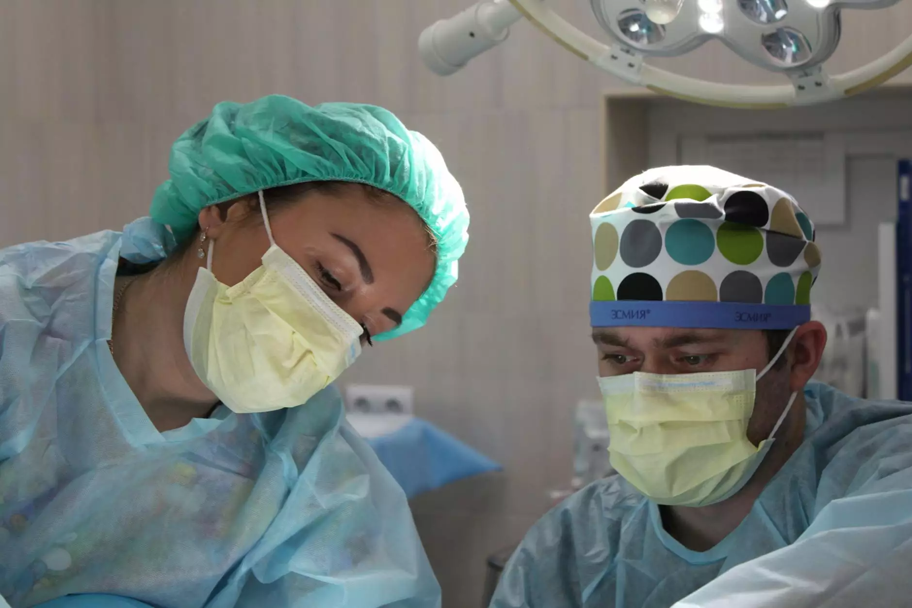
The diagnostic hysteroscopy procedure itself is performed in a clinical setting, usually in an outpatient facility. Here’s a breakdown of what patients can expect:
1. Anesthesia
Patients may receive local anesthesia, sedation, or general anesthesia, depending on individual circumstances and physician preference.
2. Insertion of the Hysteroscope
The doctor carefully inserts the hysteroscope into the vagina, through the cervix, and into the uterus. The hysteroscope has a light and camera, allowing the doctor to visualize the uterine lining.
3. Fluid Distention
A sterile fluid (saline or other solutions) is introduced into the uterine cavity to expand it, enabling a clearer view of the uterus.
4. Examination and Possible Interventions
The doctor thoroughly examines the uterine walls and cavity. If abnormal growths or issues are detected, minor surgery such as polypectomy or biopsy can be performed at this time.
5. Conclusion of the Procedure
The hysteroscope is carefully removed, and the patient is monitored for a short period before being discharged.
Recovery After Diagnostic Hysteroscopy
Recovery from a diagnostic hysteroscopy is generally quick. Here’s what to expect after the procedure:
- Rest: Most patients can resume light activities within a day.
- Post-Procedure Discomfort: Some mild cramping and spotting may occur, which usually resolves quickly.
- Follow-Up: A follow-up appointment is often scheduled to discuss findings and any necessary treatment plans.
Potential Risks and Considerations
As with any procedure, there are potential risks involved with diagnostic hysteroscopy. These may include:
- Infection: Although rare, there is a possibility of developing an infection.
- Uterine Perforation: In exceptionally rare circumstances, the hysteroscope could perforate the uterine wall.
- Bleeding: Mild bleeding can occur post-procedure but should be monitored.
- Anesthesia Complications: There are inherent risks related to anesthesia that should be discussed with the provider.
Conclusion
In summary, the diagnostic hysteroscopy procedure represents a significant advancement in the field of women's health. By allowing for direct visualization and treatment of uterine abnormalities, it empowers healthcare providers to offer tailored care and improve patient outcomes. If you are experiencing symptoms that may warrant this procedure, do not hesitate to consult with a qualified professional at a reputable clinic like Dr. Seckin’s clinic. Understanding your health is critical, and through procedures like hysteroscopy, women can take proactive steps in managing their reproductive health.
With the insights and benefits provided by diagnostic hysteroscopy, patients can look forward to more informed decisions about their gynecological health. Always consult with your doctor to understand the most effective treatment options available to you.


